Advancing AI for medicine
Weekdays find University of Dayton Research Institute sensors scientist Barath Narayanan ’13 working on sponsored research in artificial intelligence for commercial applications, or in the classroom, teaching electrical and computer engineering to UD students. But evenings and weekends, Narayanan pursues his principal passion — advancing research in AI to help doctors diagnose and treat patients more quickly.

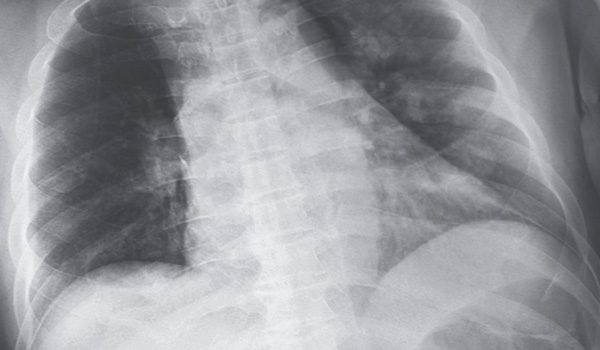
In the last few years, Narayanan has developed a number of AI-based software systems that use a machine learning or deep learning architecture to identify illness such as lung cancer and pneumonia on medical images. So when a set of chest X-rays from patients with COVID-19 became available for research just days into the pandemic, Narayanan quickly shifted focus.
Within a matter of hours, he had tailored his software to correctly classify the X-rays as having, or not having, COVID-19 in a matter of seconds and with 98 percent accuracy.
Enter Srikanth Kodeboyina ’11, who learned about Narayanan’s research from a blog entry the scientist posted just a few days after his trial success. Within three days, Kodeboyina held an exclusive license to the technology, facilitated in record time by the University’s Technology Partnerships Office. And just two weeks from the day Narayanan successfully developed COVID-19 detection software, Kodeboyina had filed a provisional patent and for FDA approval that, when granted, will allow him to bring the technology to market.
Both men were driven by the same mission: Finding a way to rapidly provide another tool in the fight against COVID-19. “Right now one of the most pressing needs on the planet is addressing the COVID-19 crisis,” Kodeboyina said. “Artificial intelligence can be an important solution to support the health care industry in its fight to mitigate the impact of the disease, and we are on the leading edge of developing that technology.”
 For Narayanan, COVID-19 is yet another disease in a long list of illnesses that could be better managed with faster diagnosis. So most of his free time — according to his wife and occasional research partner, Priyamvada Davuluru ’15— is spent in front of a computer, writing code, testing algorithms or searching for available medical images. To date, he has developed software systems that successfully detect — with 92 to 99 percent accuracy — lung and breast cancers, malaria, brain tumors, tuberculosis, diabetic retinopathy, pneumonia and COVID-19 on chest X-rays, CT scans, blood smear slides or eye scans.
He has also published three peer-reviewed journal papers and nine conference papers on his medical imaging research, with three additional papers recently accepted for publication. He traveled to Texas in February to discuss his most recent paper, on deep learning architecture for pneumonia detection, published at the International Society for Optics and Photonics’ medical imaging conference in Houston.
Narayanan, who holds master’s and doctoral degrees in electrical engineering from UD, says he’s driven by a desire to use his education and experience for the common good.
“I enjoyed the image processing and machine learning aspects of my graduate research and decided to use the field to improve the quality of human life in some way. Computer-aided diagnosis seemed a good platform to explore for that purpose,” he said.
 He targeted lung cancer for his doctoral dissertation on computer-aided detection not only for its prevalence as the leading cause of cancer deaths but also for the drastic increase in survival rates among patients who are diagnosed in early stages — 57.4%, compared with the 5.2% survival rate of patients who aren’t diagnosed until their cancer has metastasized, according to the National Cancer Institute.
“Finding ways to support doctors in early diagnosis of diseases is critical to providing the best treatments and outcomes.”
“Finding ways to support doctors in early diagnosis of diseases is critical to providing the best treatments and outcomes,” Narayanan said, confessing that part of his motivation is personal; several members of his close circle of family and friends have been affected by late diagnosis of illness.
“Software-based diagnostic tools can help reduce human error and serve as a valuable, virtual second opinion for medical professionals, especially in parts of the world where medical teams are short-staffed. With additional research, these technologies can be fine-tuned to detect even the slightest anomalies on images — those that are difficult to see with the human eye — helping doctors diagnose and treat patients more quickly.”
While advancements in computer-aided detection for medical imaging applications have been significant in recent years, Narayanan said the health care industry has been slow to employ the technology in patient care.
“Physicians are not ready to fully trust deep learning models because data scientists have not yet been able to demonstrate the process behind the system’s decisions,” he said.
Narayanan, who is working to change that, explains the challenge.
In artificial intelligence for visual applications, scientists provide software systems with input in the form of images that are labeled for content. For example, images of birds will not only be labeled generically as birds, but its eyes, beaks, tails and other characteristics will also be labeled as such, allowing the system to familiarize itself with bird components. With increased exposure to diverse avian imagery, the software will learn to look for those characteristics and, ultimately, independently identify images of birds that have not been labeled in any way.
When using a deep-learning system to classify images, however, the system’s training stage includes input with only generic labels, but no instructions on what to identify within the images.
“We feed the system multiple images and allow it to identify patterns it notes over time,” Narayanan said. “The more input it receives, the more robust the system will be in accurately identifying the image, be it a bird, a car, or a chest X-ray showing pneumonia, lung cancer or COVID.”
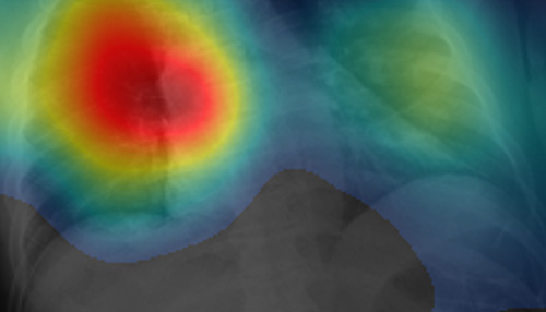
Because deep learning algorithms learn to make decisions about what is contained in an image independently, data scientists have been working to understand the mechanism behind the systems’ decision-making process, he said: “We must be able to explain why this technology is reliable before it is widely accepted within the medical community.”
Narayanan has been working to do that by programming his software to highlight “regions of interest” in red on each image, indicating where it detects anomalies that cause it to classify the image as showing a particular disease. These can be compared to a radiologist’s findings to validate the accuracy of the software as well as to support the diagnosis.
“Determining and demonstrating the region of interest that contributes to a system’s decision-making process will help data science experts better understand our systems, which in turn will help us build trust among the clinicians and patients who can greatly benefit from this technology,” he said. “It is also important that additional research to validate the technology includes a more diverse range of patient cases, because medical imaging results may be influenced by many factors, including the type of equipment being used or even the country in which patients are located.”
While Narayanan is hoping to find funding that will enable him to further develop AI tools for the medical field, he said he will continue to work on his own time to help bring them to the health care industry. He also engages UD engineering students in in his research, collaborating with them and former doctoral adviser Russell Hardie, professor of electrical and computer engineering, to develop software tools for diagnostic systems.
Hardie, who describes his young colleague as having an “oversized kindness about him and generosity of spirit,” said Narayanan is not only making important contributions that will help transition computer-aided detection technology to clinical practice, he’s also helping the next generation of researchers do the same.
“Barath is driven by the desire to harness his skills and curiosity to advance research for the common good.”
“Barath is driven by the desire to harness his skills and curiosity to advance research for the common good, as well as by his desire to connect with students,” Hardie said. “He has designed innovative class projects that leverage his wealth of knowledge in medical imaging and machine learning and captivate students, providing them with valuable collaborative experience. Our department is fortunate to have such an energetic, hard-working and generous individual in our ranks.”